
7 Best PEEK Interference Screws for ACL Reconstruction

ACL reconstruction depends on secure graft fixation, and the interference screw is a small part with a demanding job. In a 21-year population study from Olmsted County, Minnesota, Sanders and colleagues reported 68.6 ACL tears and 44.4 reconstructions per 100,000 person-years. These figures describe one regional population, not a worldwide rate. Small parts matter. A Peek Interference Screw compresses the graft against the bone tunnel, so material, diameter, length, and thread design can affect the fixation approach. PEEK is radiolucent, which can make postoperative imaging easier to interpret than imaging around some metal implants. But the screw may be less visible on standard radiographs, and product details still matter.

This guide compares seven PEEK interference screws used in ACL reconstruction, focusing on practical features rather than claiming one universal winner. The AAOS Management of ACL Injuries Clinical Practice Guideline (2022) addresses reconstruction decisions and meniscal risk; it does not establish one screw as best for every patient. That distinction matters. Graft type, tunnel dimensions, bone quality, surgical technique, and the device’s approved instructions all shape suitability. We consider design and handling details clinicians can verify, including available sizes, insertion features, and imaging considerations. Manufacturer specifications can help narrow options, but they are not the same as independent clinical evidence. Some comparisons remain imperfect because head-to-head studies are limited. A careful shortlist should make that uncertainty visible, not hide it.
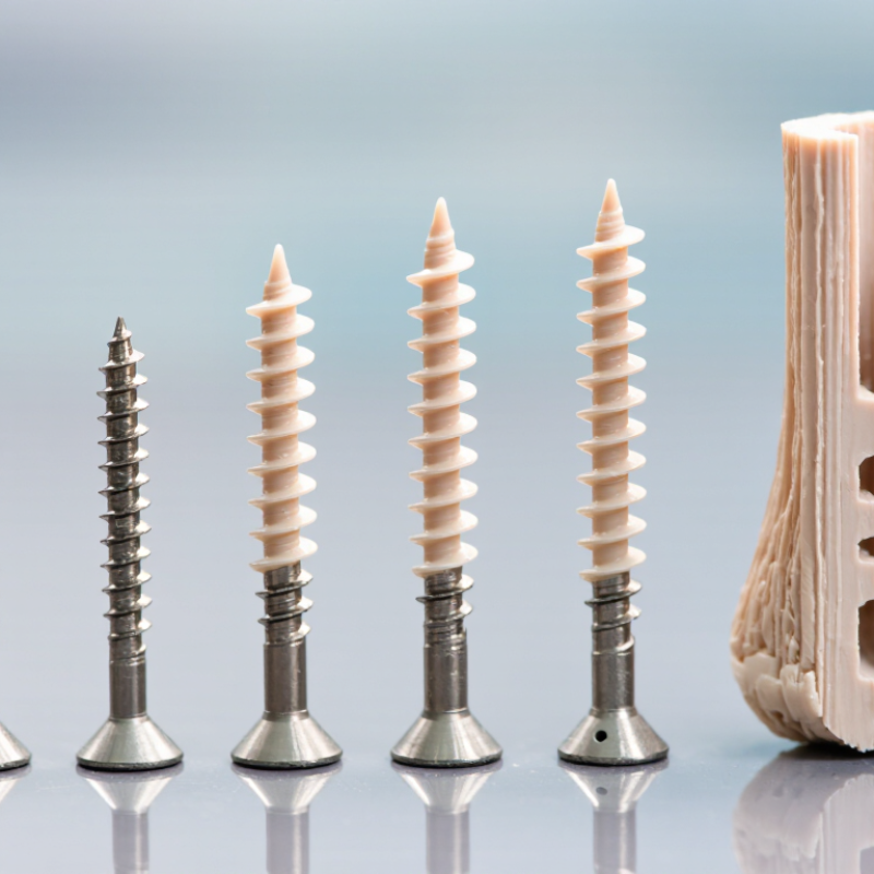
7 Best PEEK Interference Screws for ACL Reconstruction

PEEK Basics: ASTM F2026 Implant-Grade Material and ACL Screw Design

PEEK, or polyether ether ketone, is a high-performance polymer used in some implant applications. ASTM F2026 specifies requirements for PEEK polymers intended for surgical implants. It is a material standard, not a guarantee that every finished screw performs identically. Documentation should identify the material grade and relevant testing. Details matter.

For ACL reconstruction, an interference screw presses the graft against the bone tunnel wall. PEEK’s radiolucency can make the screw less conspicuous on standard X-rays than metal implants, while its shape remains visible through other imaging methods. Thread depth, pitch, and core diameter influence how the screw engages bone and compresses the graft. A blunt or tapered tip may also affect insertion, depending on the tunnel and surgical technique. Small design changes can matter.

Material choice is only part of the assessment. The screw’s dimensions, surface finish, insertion driver, and resistance to cracking under load all deserve review. Surgeons also weigh bone quality, graft type, and the planned fixation method. A datasheet cannot replace that judgment. Nor should “implant-grade” be read as proof of a particular clinical result. In practice, a screw that looks ideal on paper may feel different during insertion; that gap deserves careful testing and reflection.

PEEK Basics for ACL Interference Screws

Representative elastic modulus comparison of implant-grade PEEK, cortical bone, and titanium alloy.

PEEK’s modulus is closer to bone than titanium alloy’s in these representative comparisons. Actual properties vary with material grade, bone site, and test method. ASTM F2026 covers implant-grade PEEK material; it does not specify ACL screw dimensions or guarantee clinical performance.

Screen Seven Options by Diameter (mm), Length (mm), Thread, and Graft Type

7 Best PEEK Interference Screws for ACL Reconstruction

Screen seven practical options by diameter, length, thread, and graft type: 7 × 20 mm fine-thread for smaller hamstring grafts; 8 × 20 mm fine-thread for compact soft-tissue grafts; 8 × 25 mm coarse-thread for longer tunnel engagement; 9 × 20 mm for selected bone–patellar tendon–bone grafts; 9 × 25 mm for larger graft-tunnel combinations; 10 × 25 mm coarse-thread for broader grafts; and 10 × 30 mm where tunnel length allows. These are screening examples, not universal prescriptions. Confirm tunnel measurements, graft dimensions, and the device’s instructions for use.

Thread geometry affects insertion and graft compression. Diameter should match the tunnel and graft without forcing insertion. PEEK is radiolucent, which can help postoperative imaging, but it does not make the implant invisible on every scan. The AAOS 2022 ACL guideline discusses graft choice, while Scandinavian Knee Ligament Register annual reports track graft and revision outcomes. Those datasets inform clinical context; they do not establish one ideal screw size. A tidy size chart can still mislead.

Tip: Measure the prepared tunnel and graft directly. Check thread design and length against the surgeon’s plan. If the fit feels uncertain, pause and reassess rather than relying on a size label alone.

Compare Bench Data: Pullout Strength (N), Insertion Torque (N·cm), and Failure Mode

For seven PEEK interference screws, compare measured pullout strength, insertion torque, and failure mode under the same test conditions. Peer-reviewed ACL fixation studies show that results depend on bone density, graft type, screw diameter, and insertion depth. A pullout value in newtons is meaningful only beside those details. ASTM F543 describes mechanical testing for metallic bone screws; it does not set a PEEK-specific ACL fixation threshold. That distinction matters. A neat ranking can hide mismatched methods.

Insertion torque, reported in N·cm, helps show how much rotational force seating the screw required. Some studies report torque in N·m; multiply by 100 to convert to N·cm. Higher torque is not automatically better. It may reflect tighter graft compression, but also dense bone or difficult insertion. Record whether failure came from graft slippage, screw breakage, or bone damage. These modes tell different stories, even when pullout loads look similar. Bench results are useful, but they do not predict every patient outcome.

Tip: Ask for the full test protocol, sample size, and variability, not just the peak number. Compare like with like. If methods differ, say so plainly; the ranking may be less certain than it looks.

Assess Clinical Evidence: Follow-Up (months), Revision Rate (%), and Imaging

For seven PEEK interference screws, compare evidence by follow-up time, revision rate, and imaging—not by material claims alone. Scandinavian knee-ligament registry reports place ACL revision rates in the low single digits, roughly 3–5% at five years across fixation methods. That is useful context, not a PEEK-specific result. A screw comparison should state its patient count, graft type, and revision definition beside every percentage.

Clinical studies often have modest sample sizes and follow-up measured in months or a few years. Read each time point carefully: a 24-month result cannot establish long-term durability. Revision rates also need denominators; “two revisions” means little without knowing whether the study followed 40 patients or 400. The Swedish National Knee Ligament Register tracks revision over time, but pooled registry figures do not isolate PEEK screw performance. Important.

Imaging adds another layer. PEEK is radiolucent, so MRI can show the graft and tunnel with less implant obstruction than metal fixation. Radiographs or CT may help assess tunnel widening, but imaging alone cannot prove secure graft integration. Compare the imaging method, timing, and reported findings across studies. The evidence is uneven, and I would hesitate to rank seven options when follow-up and revision reporting differ so much.

7 Best PEEK Interference Screws for ACL Reconstruction - Assess Clinical Evidence: Follow-Up (months), Revision Rate (%), and Imaging

Evidence note: This table does not rank or identify commercial products. Published clinical results are not sufficiently standardized to provide reliable, comparable follow-up durations or revision rates for each category below. “Not established” means a defensible category-specific estimate cannot be given here; it does not mean that no clinical studies exist.

ACL reconstruction context Follow-up (months) Revision rate (%) Imaging considerations Evidence interpretation
Soft-tissue hamstring graft, tibial fixation Not established as a pooled, screw-specific value Not established as a pooled, screw-specific value PEEK is radiolucent; tunnel and graft assessment generally relies on the surrounding bone and graft rather than direct screw visibility on plain radiographs. Outcomes depend on graft, tunnel, surgical technique, and study follow-up; avoid attributing results to screw material alone.
Soft-tissue hamstring graft, femoral fixation Not established as a pooled, screw-specific value Not established as a pooled, screw-specific value PEEK typically produces less susceptibility artifact than many metallic implants, but MRI appearance depends on the scan protocol and any radiopaque marker. Femoral fixation method and tunnel position are important potential confounders when comparing clinical series.
Bone–patellar tendon–bone (BPTB) graft, tibial fixation Not established as a pooled, screw-specific value Not established as a pooled, screw-specific value Radiolucency can make the screw itself difficult to distinguish on radiographs; assess bone tunnels and bone-block position using appropriate imaging. BPTB results should not be directly compared with soft-tissue graft results without accounting for graft type and fixation protocol.
BPTB graft, femoral fixation Not established as a pooled, screw-specific value Not established as a pooled, screw-specific value PEEK is generally radiolucent; MRI can evaluate surrounding tissues, while CT may help assess tunnel and bone-block position when clinically indicated. Imaging findings are not, by themselves, a measure of clinical success or a reason for revision.
Primary ACL reconstruction, mixed graft types No single comparable duration; report each study’s actual follow-up No single comparable rate; definitions and follow-up periods vary PEEK is radiolucent and is usually less artifact-producing on MRI than metal; some screw designs may include a radiopaque marker. A valid comparison requires the same revision definition, follow-up period, graft type, and patient population.
Revision ACL reconstruction Not established as a pooled, screw-specific value Not established as a pooled, screw-specific value CT is commonly useful for evaluating tunnel position and bone stock; PEEK itself may be inconspicuous, so review the full imaging study. Revision cases differ substantially from primary reconstruction and should be analyzed separately.
Cross-study imaging and safety assessment Report the imaging time point for each study Not an imaging-derived measure Radiolucency can improve visualization of nearby anatomy compared with metal, but may reduce direct visibility of the screw. Confirm implant-specific MRI conditions and marker details. Imaging modality, acquisition protocol, and radiographic endpoints should be stated before comparing studies.

Select by Tunnel Size (mm), Graft Compatibility, and Manufacturer’s IFU

Choosing among seven PEEK interference screw options starts with tunnel measurements, not a product label. Confirm the prepared tunnel’s diameter and length, then compare them with the screw’s stated dimensions and the manufacturer’s IFU. A close fit matters. Oversizing may affect graft compression or insertion; undersizing may provide less fixation than intended. These decisions belong to the operating surgeon, using the patient’s anatomy and the surgical technique.

Graft compatibility narrows the field. Hamstring soft-tissue grafts and bone–patellar tendon–bone grafts have different shapes and fixation needs. Check whether the IFU specifically covers the planned graft type, screw design, and insertion instruments. Thread profile and tip geometry can influence how smoothly a screw advances beside the graft. Small details count. A screw that appears suitable by diameter may still be a poor match for a particular tunnel or graft construct.

Compare the seven candidates by available diameters, lengths, graft indications, and required tools—not by headline claims alone. Read the current IFU for placement guidance, contraindications, and any limits on reuse or imaging. PEEK is not a guarantee of better outcomes. Evidence and results vary by technique and patient. One honest limitation: published comparisons may not answer every sizing question. When measurements sit between options, document the rationale and follow the surgeon’s established protocol.